Left Shoulder Anatomy Diagram / The Shoulder Joint - Click on a view to review its anatomy.. I have hiked this most beautiful place 15 times and dr. Webmd's shoulder anatomy page provides an image of the parts of the shoulder and describes its function, shoulder problems, and more. Month ago i fell on my left shoulder while on a bush walk, hard fall. Did not undergo a x ray or. Shoulder anatomy is an important part of understanding the treatment of the rotator cuff syndrome.
Webmd's shoulder anatomy page provides an image of the parts of the shoulder and describes its function, shoulder problems, and more. Learn vocabulary, terms and more with flashcards, games and other study tools. To keep things simple, we can divide the shoulder into layers. Normal anatomy, variants and checklist. Human body anatomy human anatomy and physiology body muscle anatomy anatomy study anatomy reference muscle diagram psoas release muscular system medical anatomy.
Left Arm Muscle Model Labeled Google Search Arm Muscle Anatomy Body Anatomy Human Muscle Anatomy from i.pinimg.com Required fields are marked *. Learn vocabulary, terms and more with flashcards, games and other study tools. Start studying shoulder anatomy diagram. Robin smithuis and henk jan van der woude. Understanding how the different layers of the shoulder are built and connected can help you understand how the shoulder works, how it can be injured, and how challenging recovery can be. Month ago i fell on my left shoulder while on a bush walk, hard fall. Bone, then ligaments of the joint capsule, with tendons and muscles on top. When the leg is stretched out, the knee joint is placed on a straight line with the hip and ankle (left).
Normal anatomy, variants and checklist.
Normal anatomy, variants and checklist. I have hiked this most beautiful place 15 times and dr. Radiologists primarily perform shoulder imaging to assess injuries within the shoulder joint. Enjoy the videos and music you love, upload original content, and share it all with friends, family, and the world on youtube. The anatomy of the provides the strength and functionality of the upper body. The arm bone (humerus), the shoulder blade. Use the mouse scroll wheel to move the images up and down alternatively use the tiny arrows (>>) on both side of the image to move the images. The sagittal suture is the line where the right and left parietal bone are in contact. Posted on december 13, 2018december 12, 2018. Related posts of diagram of shoulder muscles and tendons. Start studying shoulder anatomy diagram. 7 draw labelled diagram showing the relations of shoulder joint. Understanding how the different layers of the shoulder are built and connected can help you understand how the shoulder works, how it can be injured, and how challenging recovery can be.
I have hiked this most beautiful place 15 times and dr. Posted on december 13, 2018december 12, 2018. The shoulder is one of the largest and most complex joints in the body. The arm bone (humerus), the shoulder blade. The shoulder joint is the connection between the chest and the upper extremity.
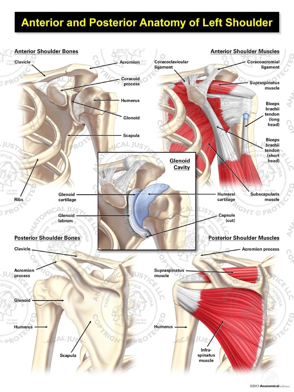
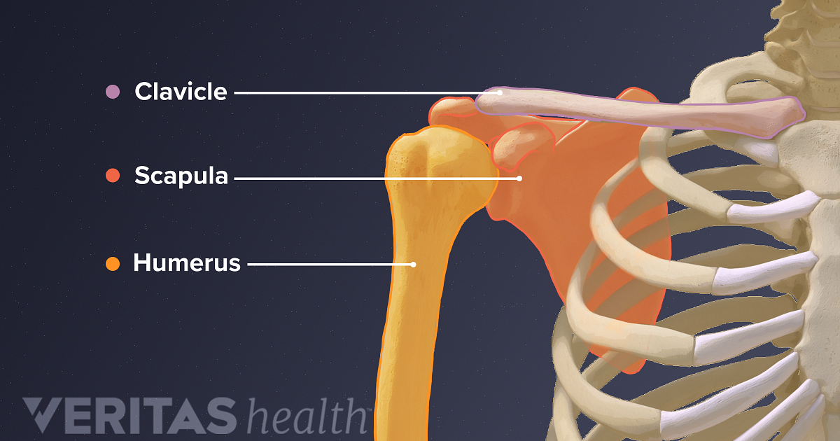
Shoulder Anatomy Posterior Anatomy Drawing Diagram from anatomicaljustice.com Start studying shoulder anatomy diagram. This webpage presents the anatomical structures found on shoulder mri. Understanding how the different layers of the shoulder are built and connected can help you understand how the shoulder works, how it can be injured, and how challenging recovery can be. The shoulder can counteract an extreme impact but is also vulnerable to to a range of pathologies due to inactivity, overuse and trauma. When the leg is stretched out, the knee joint is placed on a straight line with the hip and ankle (left). 7 draw labelled diagram showing the relations of shoulder joint. The sagittal suture is the line where the right and left parietal bone are in contact. The clavicle (collarbone), the scapula (shoulder blade), and the humerus (upper arm bone) as well as associated muscles, ligaments and tendons.
When the leg is stretched out, the knee joint is placed on a straight line with the hip and ankle (left).
When the leg is stretched out, the knee joint is placed on a straight line with the hip and ankle (left). The shoulder is one of the largest and most complex joints in the body. The anatomy of the provides the strength and functionality of the upper body. The arm bone (humerus), the shoulder blade. The human shoulder is made up of three bones: Bone, then ligaments of the joint capsule, with tendons and muscles on top. The shoulder joint is formed where the humerus (upper arm bone) fits into the scapula. Editor · aug 6, 2017 ·. Your email address will not be published. The sagittal suture is the line where the right and left parietal bone are in contact. Sechrest, md narrates an animated tutorial on the basic anatomy of the shoulder. I have hiked this most beautiful place 15 times and dr. Pain is common in athletes and the general public.
Start studying shoulder anatomy diagram. This flow diagram provides an aid to anatomy, shoulder and upper limb, hand cutaneous innervation. statpearls internet. Learn vocabulary, terms and more with flashcards, games and other study tools. Humbert's joint implant of my left hip enabled me to continue my love of hiking my favorite place. 7 draw labelled diagram showing the relations of shoulder joint.
Guide To Shoulder Anatomy from embed.widencdn.net The shoulder joint is encapsulated by a group of muscles and ligaments called the rotator cuff. Humbert's joint implant of my left hip enabled me to continue my love of hiking my favorite place. To begin with, it's slightly curved down, but in tension the shoulders tense up and the curve can itself turn up and look higher. The human shoulder is made up of three bones: As the disease progresses, night pain becomes more common. The anatomy of the provides the strength and functionality of the upper body. Movements of the human shoulder represent the result of a complex dynamic interplay of structural bony anatomy and biomechanics, static a thorough understanding of the functional anatomy of the shoulder provides the clinician with a foundation for caring for athletes with shoulder injuries. As a ball and socket synovial.
Ac joint is a diathrodial joint with a fibrocartilaginous disk.
8 name the arteries and the nerves that supply shoulder leave a reply cancel reply. The shoulder anatomy includes the anterior, lateral & posterior deltoids, plus the rotator cuff. The anatomy of the provides the strength and functionality of the upper body. Human body anatomy human anatomy and physiology body muscle anatomy anatomy study anatomy reference muscle diagram psoas release muscular system medical anatomy. Leave a reply cancel reply. The shoulder can counteract an extreme impact but is also vulnerable to to a range of pathologies due to inactivity, overuse and trauma. To keep things simple, we can divide the shoulder into layers. Bone, then ligaments of the joint capsule, with tendons and muscles on top. Starting with what is deepest, it goes: Humbert's joint implant of my left hip enabled me to continue my love of hiking my favorite place. Learning to see and draw energy. Related posts of diagram of shoulder muscles and tendons. This acts as the bony framework by which the muscles of the chest, upper back and shoulder connect the upper limb to the trunk of the body and control it's movements.the clavicle connects to the sternum via the.
This mri shoulder axial cross sectional anatomy tool is absolutely free to use shoulder anatomy diagram. After leaving the plexus it divides into the anterior and posterior branch.


0 Komentar